Tuesday, 24 September 2024

From Strength to Recovery | The Essential Role of Kinematic Chain Exercises in Physiotherapy | Blog by CB Physiotherapy, Active Healing for Pain Free Life.


In physiotherapy, the ultimate goal is to restore function, improve strength, mobility, and range of motion, and minimize pain for individuals recovering from injury or surgery. A critical part of achieving this involves the use of specific exercise techniques, particularly open and closed kinematic chain exercises. These exercises form the backbone of rehabilitation programs, helping patients progress safely and effectively toward full recovery.
This blog will explore what these exercises entail, their respective advantages, and how they are used in physiotherapy treatment.

Understanding Kinematic Chains: Open vs. Closed

Kinematic chain exercises refer to movements that involve multiple joints and muscles working together in a sequence. These exercises are broadly categorized into two types: open and closed kinematic (or kinetic) chain exercises.

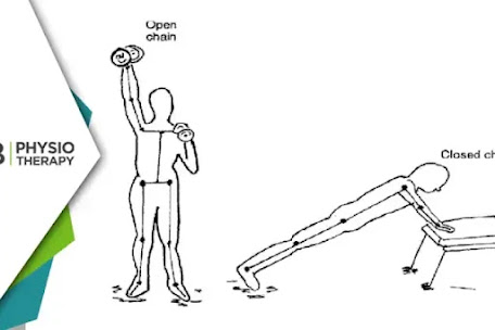
Open Kinematic Chain Exercises In an open kinematic chain (OKC) exercise, the distal (farthest) part of the limb moves freely in space, without a fixed point of contact with the surface. For example, when you kick a ball or extend your leg while seated, the foot (distal limb) is free to move, and the rest of the body remains relatively stationary. OKC exercises typically isolate a specific muscle group or joint, focusing on improving strength and range of motion in a controlled and targeted manner.

Examples of Open Kinematic Chain Exercises:
1. Leg extensions
2. Bicep curls
3. Seated knee extensions
4. Shoulder presses

Closed Kinematic Chain Exercises In contrast, closed kinematic chain (CKC) exercises involve movements where the distal limb is fixed or in contact with a surface (ground or another stable object), and the body moves in relation to it. For example, during a squat, the feet remain in contact with the ground while the body moves. CKC exercises tend to engage multiple muscle groups simultaneously, promoting joint stability and overall coordination.

Examples of Closed Kinematic Chain Exercises:
1. Squats
2. Push-ups
3. Lunges
4. Deadlifts

Role of Open and Closed Kinematic Chain Exercises in Physiotherapy

Both open and closed chain exercises have distinct roles in rehabilitation, offering unique benefits depending on the patient’s needs and the specific phase of treatment. The decision to incorporate either or both types depends on factors such as the type of injury, the joint involved, the stage of healing, and the goals of therapy.

1. Open Kinematic Chain Exercises: Targeted Strengthening and Range of Motion OKC exercises are ideal for isolating and strengthening specific muscles or joints. In the early stages of rehabilitation, particularly following surgery or injury, physiotherapists often prioritize OKC exercises because they place less overall stress on the joints, reducing the risk of re-injury while allowing controlled muscle activation.

a: Advantages of Open Kinematic Chain Exercises:
1. Isolation of Specific Muscles: OKC exercises are excellent for targeting individual muscles that may be weak or underused due to injury. For example, after ACL reconstruction surgery, physiotherapists may prescribe knee extensions to target the quadriceps.

2. Improved Range of Motion: These exercises are beneficial for improving the range of motion in joints like the knee or shoulder. Isolated movements help stretch and strengthen muscles around the joint, facilitating recovery of movement patterns.

3. Early-Stage Rehabilitation: OKC exercises are often used in the early stages of treatment when the patient may not yet be able to tolerate weight-bearing activities. They allow for muscle activation without placing undue strain on healing tissues.

b: Clinical Application of Open Kinematic Chain Exercises:
1. Post-Surgery Rehabilitation: After surgeries like a knee replacement or rotator cuff repair, OKC exercises are often prescribed to gradually strengthen muscles without stressing the joint.

2. Rehabilitation of Non-Weight Bearing Injuries: OKC exercises are critical in scenarios where weight-bearing is not advised, such as in certain fractures or post-operative conditions.

2. Closed Kinematic Chain Exercises: Functional Strength and Joint Stability CKC exercises are more functional in nature and closely mimic the movements that patients perform in their daily activities. These exercises recruit multiple muscle groups, promoting coordination, balance, and joint stability. In physiotherapy, CKC exercises are particularly beneficial for improving proprioception (awareness of joint position), which is often impaired after injury.

A: Advantages of Closed Kinematic Chain Exercises:
1: Joint Stability: CKC exercises help stabilize joints by engaging multiple muscles around the joint. For example, during a squat, the quads, hamstrings, and glutes all work together to provide stability to the knee joint, reducing the risk of injury.

2: Functional Movement Patterns: These exercises mimic everyday movements, making them highly functional. For example, squats and lunges resemble activities like standing up from a chair or climbing stairs, helping patients regain the ability to perform daily tasks.

3: Improved Proprioception: Because CKC exercises involve weight-bearing movements, they are excellent for enhancing proprioception and balance. This is particularly important for patients recovering from lower extremity injuries, such as ankle sprains or knee ligament tears.

4: Multi-Joint Coordination: CKC exercises engage multiple joints and muscle groups simultaneously, improving coordination and helping patients restore full-body strength and stability.

B: Clinical Application of Closed Kinematic Chain Exercises:
1: Late-Stage Rehabilitation: As patients progress through their rehabilitation program, CKC exercises are introduced to improve functional strength and prepare them for the demands of daily life or sports activities. For example, after knee surgery, physiotherapists may prescribe squats and lunges to strengthen the lower limbs and improve overall stability.

2: Prevention of Re-Injury: CKC exercises are crucial for preventing future injuries by reinforcing joint stability and muscle coordination. This is particularly beneficial for athletes returning to sports after injury.

Combining Open and Closed Kinematic Chain Exercises in Physiotherapy

For optimal results in physiotherapy, combining both OKC and CKC exercises is often the most effective approach. The integration of these exercises allows for comprehensive rehabilitation, addressing both the specific muscle weakness and the overall functional stability of the body.

1: Early-Stage Rehabilitation:
In the initial phases of rehabilitation, OKC exercises are typically prioritized to isolate and strengthen specific muscles. For example, after an ACL reconstruction, the physiotherapist may focus on leg extensions to target the quadriceps without placing too much stress on the healing knee joint.

2: Mid-to-Late Stage Rehabilitation:
As the patient progresses, CKC exercises like squats and lunges are introduced to promote joint stability, functional strength, and proprioception. This prepares the patient for the demands of everyday activities and sports, ensuring that the joint can handle complex, multi-joint movements.

By incorporating both open and closed chain exercises, physiotherapists can tailor rehabilitation programs to meet the individual needs of each patient, ensuring a safe and effective recovery. Understanding the importance and proper application of these exercises allows patients to regain strength, mobility, and function more effectively, ultimately improving their quality of life.
Hill-Sachs Lesion | How Physiotherapy Can Reduce the Risk of Recurrent Shoulder Dislocations? | Blog by CB Physiotherapy, Active Healing for Pain Free Life.


 The shoulder joint, being one of the most mobile joints in the human body, is vulnerable to dislocation and associated injuries. One of the common complications of shoulder dislocation is a Hill-Sachs lesion-a compression fracture or indentation on the posterolateral aspect of the humeral head. This lesion is often associated with recurrent shoulder dislocations, particularly in active or younger individuals. Understanding the biomechanics of this condition and how physiotherapy can manage it is crucial for effective rehabilitation and prevention of further dislocations.

Understanding a Hill-Sachs Lesion

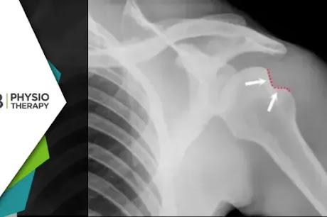
A Hill-Sachs lesion occurs when the humeral head, the ball of the shoulder joint, forcefully impacts the edge of the glenoid cavity (socket) during a shoulder dislocation. The most common type of shoulder dislocation is anterior dislocation, where the humeral head slips out of the socket in a forward direction. As the humeral head is driven against the glenoid rim, it creates a dent or compression fracture on the humerus, which is referred to as a Hill-Sachs lesion.

Link Between Hill-Sachs Lesion and Recurrent Shoulder Dislocation

When a Hill-Sachs lesion is present, it alters the shape of the humeral head, making it more prone to catching on the rim of the glenoid, particularly during movements that involve external rotation and abduction (raising the arm sideways). This biomechanical alteration increases the risk of the shoulder slipping out of place again, resulting in chronic shoulder instability.

In more severe cases, a Bankart lesion-damage to the anterior labrum of the shoulder-may accompany the Hill-Sachs lesion, further destabilizing the joint. The combination of these injuries can lead to recurrent shoulder dislocations, especially in athletes who participate in contact sports or individuals who frequently engage in overhead activities.

The symptoms of a Hill-Sachs lesion may vary depending on its severity and the frequency of shoulder dislocations. Common signs include shoulder pain that worsens with movement, sense of instability or weakness in the shoulder, particularly during overhead movements, limited range of motion, especially in external rotation and abduction, recurrent dislocations, sometimes with minimal force, creaking or clicking sounds during shoulder movements. If left untreated, these symptoms can progressively worsen, significantly impacting a person’s daily life and activities.

Physiotherapy in the Management of Hill-Sachs Lesion and Shoulder Instability Physiotherapy plays a pivotal role in managing both the immediate and long-term effects of a Hill-Sachs lesion, particularly in preventing recurrent shoulder dislocations. The primary goals of physiotherapy are to:
1: Restore shoulder strength and stability.
2: Improve joint proprioception.
3: Enhance functional movement patterns.
4: Reduce the risk of future dislocations.

1. Initial Phase: Pain and Inflammation Management
Following a shoulder dislocation and diagnosis of a Hill-Sachs lesion, the initial focus of physiotherapy is to control pain and inflammation. Modalities such as ice therapy, electrical stimulation, and manual therapy may be used to manage pain and promote healing of the injured tissues. Immobilization of the shoulder in a sling may be recommended for a short period, but prolonged immobilization should be avoided to prevent stiffness and muscle weakness.

2. Restoring Range of Motion (ROM)
Once pain and inflammation are under control, the next phase involves gradually restoring range of motion in the shoulder joint. This is done through passive and active-assisted exercises that focus on gently stretching the muscles and soft tissues surrounding the shoulder. Some examples of ROM exercises include:
1: Pendulum exercises: These involve gently swinging the arm in a circular motion while leaning forward, which helps increase joint mobility without putting stress on the shoulder.
2: Passive shoulder flexion and external rotation: The therapist may assist the patient in gently raising the arm overhead or rotating the arm outward.
It’s crucial to avoid any activities that place excessive strain on the shoulder, such as lifting heavy objects or performing sudden overhead movements, as this could lead to further dislocation.

3. Strengthening Shoulder Muscles
Once full or near-full range of motion is achieved, the focus shifts to strengthening the muscles that support and stabilize the shoulder joint. The rotator cuff muscles (supraspinatus, infraspinatus, teres minor, and subscapularis) play a critical role in maintaining shoulder stability and preventing dislocations.
Strengthening exercises should be progressive and target both the rotator cuff and scapular stabilizers to ensure proper shoulder mechanics. Some effective strengthening exercises include:
1: Isometric shoulder exercises: These involve contracting the shoulder muscles without moving the joint, helping to build strength without aggravating the lesion.
2: External and internal rotation exercises: Using resistance bands or light weights, patients can gradually build strength in the rotator cuff muscles, particularly the infraspinatus and subscapularis.
3: Scapular stabilization exercises: Strengthening the muscles around the scapula (shoulder blade) is important for overall shoulder stability. Exercises such as scapular retractions and wall push-ups can be helpful.

4. Enhancing Proprioception and Neuromuscular Control
Proprioception refers to the body’s ability to sense its position in space. In the case of shoulder instability, proprioception is often impaired, making it difficult for the body to detect when the shoulder is in a vulnerable position. Physiotherapy aims to improve proprioception through exercises that enhance neuromuscular control.
Balance and coordination exercises, such as using a stability ball, wobble board, or performing closed-chain exercises like weight-bearing shoulder taps, can help improve proprioception and prevent further dislocations.

5. Functional Training and Return to Activity
As strength and stability improve, the final phase of physiotherapy involves functional training that mimics the patient’s daily activities or sports-related movements. The goal is to ensure that the shoulder can safely handle the demands of daily life or athletic performance without the risk of re-injury.

In cases of severe Hill-Sachs lesions or when recurrent dislocations persist despite comprehensive physiotherapy, surgical intervention may be considered. lesions. In such cases physiotherapy is essential post-surgery to ensure a successful recovery and prevent future instability.

Is Your Butt Asleep? Discover the Role of Physiotherapy in Managing Dead Butt Syndrome | Blog by CB Physiotherapy, Active Healing for Pain Free Life.


 In today’s sedentary world, where sitting for long hours has become the norm, various health issues have arisen, one of which is Dead Butt Syndrome (DBS). Despite its somewhat humorous name, DBS is a real condition that affects many individuals, particularly those who lead inactive lifestyles or engage in repetitive movements without adequate muscle engagement. DBS is more formally known as Gluteus Medius Tendinopathy, and it primarily affects the gluteal muscles, leading to discomfort, poor posture, and even injury if left untreated.

This blog will delve into what Dead Butt Syndrome is and most importantly, how physiotherapy plays a vital role in its management and recovery.

What is Dead Butt Syndrome?

Dead Butt Syndrome refers to the weakening or underactivation of the gluteus medius muscle, one of the three primary muscles in the buttocks responsible for stabilizing the pelvis, supporting the lower back, and enabling proper hip movement. When the gluteus medius becomes underactive, it affects posture, gait, and overall body mechanics, causing strain on other muscles and joints to compensate for the weakened glutes.

One common scenario in which DBS occurs is when individuals sit for extended periods. Sitting for long hours can lead to the glutes being compressed and inactive, causing them to “forget” how to function correctly. This muscle inhibition or dysfunction leads to the muscles becoming weak or even painful, engaging in repetitive exercises like running or cycling without focusing on strengthening the gluteal muscles can also lead to an imbalance. When the glutes aren’t adequately activated, other muscles-like the hip flexors and hamstrings-take over, creating further weakness and instability, sitting with poor posture can exacerbate the issue, as slouching or leaning forward places additional strain on the hips and lower back, individuals who have suffered injuries, particularly to the lower back, hips, or knees, may also experience gluteal weakness as a secondary effect of prolonged rest or avoidance of movement.

Recognizing the signs of Dead Butt Syndrome is crucial for timely intervention. Some common symptoms include weak glutes that put extra pressure on the lower back, leading to discomfort or even chronic pain, hips may become sore or tender due to the imbalance caused by underactive gluteal muscles. with the glutes not supporting proper movement patterns, the knees often take on extra stress, leading to knee pain or injury over time, poor gluteal activation can affect posture, leading to forward tilting of the pelvis or exaggerated arching of the lower back (lordosis), general weakness in the buttocks, particularly when performing activities like climbing stairs or standing up from a seated position.

Role of Physiotherapy in Managing Dead Butt Syndrome

Physiotherapy plays a crucial role in both preventing and managing Dead Butt Syndrome. The primary goal of treatment is to strengthen the gluteal muscles, correct muscle imbalances, and restore proper movement patterns. A physiotherapist will assess the individual’s symptoms, movement, and muscle function before developing a personalized rehabilitation plan.

Here are some key physiotherapy strategies used to treat Dead Butt Syndrome:

1. Gluteal Strengthening Exercises
The cornerstone of physiotherapy for DBS is reactivating and strengthening the gluteus medius (and other gluteal muscles). A physiotherapist will prescribe targeted exercises to progressively build strength and endurance in the glutes. These exercises typically start with low-load movements and gradually increase in intensity as the patient improves.
Some of the common gluteal strengthening exercises include:
1: Clamshells: Lying on your side with knees bent, lifting and lowering the top knee while keeping the feet together.
2: Bridges: Lying on your back with knees bent, lifting your hips off the ground while squeezing your glutes.
3: Hip Thrusts: Similar to bridges but performed with the upper back resting on a bench or platform, emphasizing full hip extension.
4: Side-Lying Leg Raises: Lifting and lowering the top leg while lying on your side, targeting the outer hip muscles.
5: Squats: Focusing on proper form and ensuring glute activation during each repetition.

2. Postural and Gait Correction
A physiotherapist will assess the individual’s posture and gait to identify any compensatory patterns or imbalances. Often, poor posture and altered walking or running mechanics are contributing factors to gluteal dysfunction. Through corrective exercises and movement retraining, physiotherapy helps restore proper alignment, which reduces strain on the hips, back, and knees.

3. Pelvic Stability Work
Gluteus medius dysfunction can lead to pelvis instability, affecting the entire kinetic chain. Physiotherapy includes exercises that focus on improving pelvic stability, ensuring that the pelvis remains level during activities like walking, running, or standing. Exercises may involve balancing movements, single-leg stance work, or dynamic core strengthening to support the pelvis.

4. Manual Therapy
Physiotherapists may also use manual therapy techniques to address tightness or restrictions in the surrounding muscles, such as the hip flexors or lower back. Techniques such as myofascial releasesoft tissue mobilization, and stretching can alleviate pain, improve mobility, and encourage gluteal activation.

5. Neuromuscular Re-Education
In cases where the glutes have “forgotten” how to activate properly, neuromuscular re-education exercises may be employed. This type of therapy focuses on retraining the brain and nervous system to engage the correct muscles during movement. Physiotherapists may use techniques like electrical stimulation, biofeedback, or specific movement cues to enhance muscle activation and coordination.

6. Functional Movement Training
Functional movement training aims to incorporate the newly strengthened glutes into everyday activities and sports. A physiotherapist will design exercises that mimic real-life movements, such as squatting, lunging, or climbing stairs. The goal is to integrate gluteal activation into functional tasks, ensuring the muscles work correctly in different contexts.

7. Stretching and Flexibility Training
Tight muscles, particularly in the hip flexors and hamstrings, can exacerbate Dead Butt Syndrome. Physiotherapists often incorporate stretching routines to improve flexibility, allowing for better gluteal engagement. Regular stretching helps relieve tension, increase mobility, and promote balanced muscle function.

Dead Butt Syndrome may sound trivial, but it can significantly impact an individual’s movement, posture, and overall well-being if left unaddressed. By understanding the causes and symptoms, individuals can take proactive steps to prevent its onset, especially through regular exercise and avoiding prolonged sitting. Physiotherapy plays a crucial role in managing DBS by targeting the root of the problem-gluteal weakness and imbalance.

Understanding Women’s Back Pain | How Physiotherapy Addresses Common Conditions and Enhances Relief? | Blog by CB Physiotherapy, Active Healing for Pain Free Life.


 Back pain is a common issue that affects millions of people worldwide, with women being particularly susceptible to certain conditions. Factors such as hormonal fluctuations, pregnancy, and specific lifestyle patterns contribute to this vulnerability. This blog explores the various back pain conditions that predominantly affect women and how physiotherapy can offer effective relief and management.

Common Back Pain Conditions in Women

1: Osteoporosis-Related Fractures
Osteoporosis is a condition characterized by weakened bones, making them more prone to fractures. Women, particularly post-menopausal women, are at a higher risk of developing osteoporosis due to the decrease in estrogen levels, which plays a crucial role in maintaining bone density. Spinal fractures caused by osteoporosis can lead to chronic back pain, reduced mobility, and a significant decline in quality of life.

2: Pregnancy-Related Back Pain
Pregnancy is a joyous time but can also bring about significant physical changes that may lead to back pain. The growing uterus, hormonal changes, and weight gain shift the body’s center of gravity, increasing the strain on the back muscles and ligaments. Conditions such as sciatica, where the sciatic nerve is compressed, can cause sharp, radiating pain from the lower back to the legs.

3: Menstrual-Related Back Pain
Menstrual periods can bring about various symptoms, including lower back pain. This pain is often related to dysmenorrhea, where the uterus contracts to shed its lining, causing cramping that can radiate to the lower back. Hormonal fluctuations during the menstrual cycle can also lead to water retention and bloating, further contributing to discomfort in the back.

4: Endometriosis
Endometriosis is a condition where the tissue similar to the lining inside the uterus grows outside the uterus. This can cause chronic pelvic pain that often radiates to the lower back. Endometriosis-related back pain can be debilitating and is often accompanied by other symptoms such as fatigue, painful periods, and pain during intercourse.

5: Fibromyalgia
Fibromyalgia is a chronic pain condition that disproportionately affects women. It is characterized by widespread musculoskeletal pain, including back pain, accompanied by fatigue, sleep disturbances, and cognitive difficulties. The exact cause of fibromyalgia is not well understood, but it is believed to involve a combination of genetic, environmental, and psychological factors.

6: Ankylosing Spondylitis
Ankylosing spondylitis (AS) is a type of inflammatory arthritis that primarily affects the spine and sacroiliac joints. It is more common in men, but women who have AS often experience it differently, with more diffuse and widespread pain, including in the lower back. AS can lead to stiffness and fusion of the vertebrae, significantly impacting mobility and quality of life.

Role of Physiotherapy in Preventing Back Pain

In addition to treating existing conditions, physiotherapy also plays a vital role in preventing back pain. Regular physiotherapy sessions can help identify and address potential risk factors before they lead to chronic pain. Some of the preventive strategies include:

1: Postural Training
Poor posture is a significant contributor to back pain. Physiotherapists can assess your posture and recommend exercises and ergonomic adjustments to improve alignment and reduce strain on the back.

2: Core Strengthening
A strong core provides better support for the spine, reducing the risk of back pain. Physiotherapists can guide you through exercises that target the abdominal, back, and pelvic muscles.

3: Education
Understanding how to move safely during daily activities is crucial in preventing back pain. Physiotherapists can educate you on proper body mechanics, lifting techniques, and ergonomic practices to minimize the risk of injury.

4: Flexibility Exercises
Maintaining flexibility in the spine and surrounding muscles is essential for preventing stiffness and pain. Physiotherapists can design stretching programs to keep your muscles and joints limber.

5: Stress Management:
Chronic stress can contribute to muscle tension and back pain. Physiotherapists may incorporate relaxation techniques, breathing exercises, and mindfulness practices into your treatment plan to help manage stress and reduce pain.

Back pain is a multifaceted issue that can significantly impact a woman’s quality of life. Physiotherapy offers a holistic, personalized approach to managing and preventing back pain, addressing both the physical and emotional aspects of these conditions.

By working with a skilled physiotherapist, women can gain the tools and knowledge needed to alleviate pain, improve mobility, and enhance their overall well-being.

Managing Scheuermann’s Disease | How Physiotherapy Can Improve Spine Health and Posture? | Blog by CB Physiotherapy, Active Healing for Pain Free Life.


 Scheuermann’s Disease, also known as Scheuermann’s Kyphosis, is a condition that primarily affects adolescents and results in a forward rounding of the upper back. Named after the Danish radiologist Holger Scheuermann who first described the condition in 1921, it is considered one of the most common causes of structural kyphosis during adolescence. This blog delves into the nature of Scheuermann’s Disease and explores how physiotherapy can play a vital role in managing the condition.

What is Scheuermann’s Disease?

Scheuermann’s Disease is a growth disorder of the vertebrae, where the bones of the spine grow unevenly, with the front growing slower than the back. This uneven growth leads to a wedge-shaped deformity of the vertebrae, which causes the spine to bend forward abnormally. The condition typically affects the thoracic (upper back) region but can also occur in the lumbar (lower back) region.

The exact cause of Scheuermann’s Disease is still unknown, though it is believed to be multifactorial, involving genetic, mechanical, and possibly endocrine factors. It tends to occur more frequently in boys than girls, with symptoms often appearing between the ages of 10 and 15.

The most noticeable symptom of Scheuermann’s Disease is an abnormal curvature of the spine, typically resulting in a rounded, hunchback appearance. This kyphosis is usually rigid, meaning the spine cannot be straightened fully by posture correction alone. Other symptoms can include particularly in the area of the curvature. Pain often worsens with activity or prolonged standing or sitting. Individuals may experience stiffness in the back and a reduced range of motion, particularly in the thoracic spine. Due to the abnormal posture, muscles in the back can become fatigued more quickly, leading to overall tiredness.

In severe cases, where the curvature is extreme, the disease can lead to respiratory and cardiovascular issues due to the compression of the chest cavity.

Treatment Options for Scheuermann’s Disease

The treatment approach for Scheuermann’s Disease depends on the severity of the condition and the symptoms experienced by the patient. Treatment options range from observation and conservative management to surgical intervention in extreme cases.

Role of Physiotherapy in Managing Scheuermann’s Disease

Physiotherapy is a cornerstone in the conservative management of Scheuermann’s Disease. The primary goals of physiotherapy are to reduce pain, improve posture, enhance spinal flexibility, and strengthen the muscles supporting the spine. Below are the key components of a physiotherapy program for Scheuermann’s Disease:

1: Postural Education and Correction
A key focus of physiotherapy for Scheuermann’s Disease is teaching patients about proper posture and maintaining it. Although the kyphosis in Scheuermann’s Disease is structural and cannot be fully corrected by posture alone, improving postural awareness can help reduce pain and prevent further progression of the curvature. Physiotherapists educate patients on how to align their spine correctly and practice good posture during daily activities.

2: Stretching Exercises
Stretching exercises are designed to increase flexibility in the muscles and ligaments surrounding the spine, particularly in the thoracic region. These exercises target tight muscles, such as the pectorals (chest muscles) and hamstrings, which can contribute to the abnormal posture seen in Scheuermann’s Disease. Regular stretching helps to alleviate stiffness and improve the range of motion in the spine.

Common stretches may include:
a: Chest stretches: To open up the chest and counteract the forward pull caused by tight pectoral muscles.
b: Hamstring stretches: To alleviate tightness in the back of the thighs, which can contribute to poor posture.
c: Thoracic extension exercises: To promote the extension of the thoracic spine, counteracting the forward curvature.

3: Strengthening Exercises
Strengthening exercises are crucial in stabilizing the spine and improving the overall posture. These exercises focus on strengthening the muscles that support the spine, including the core muscles, back extensors, and scapular stabilizers. A strong core provides better support for the spine, reducing the load on the vertebrae and potentially slowing the progression of the curvature.

Examples of strengthening exercises include:
a: Core stabilization exercises: Such as planks and bridges, which strengthen the abdominal and back muscles.
b: Back extension exercises: To strengthen the muscles along the spine, particularly the erector spinae.
c: Scapular retraction exercises: To strengthen the muscles between the shoulder blades, helping to correct rounded shoulders and improve posture.

4: Manual Therapy
Manual therapy techniques, including joint mobilizations and soft tissue massage, can be beneficial in managing Scheuermann’s Disease. Joint mobilizations help to improve the mobility of the spine, particularly in areas where movement is restricted due to the kyphosis. Soft tissue massage can alleviate muscle tension, reduce pain, and improve circulation in the affected areas.

5: Breathing Exercises
In cases where Scheuermann’s Disease affects the thoracic region severely, it can impact lung function by compressing the chest cavity. Breathing exercises can be incorporated into physiotherapy to enhance lung capacity and improve respiratory function. These exercises focus on diaphragmatic breathing and expanding the rib cage, promoting better oxygen intake and reducing the risk of respiratory complications.

6: Bracing and Orthotics
In certain cases, particularly in younger patients with a more significant curvature, bracing may be recommended in conjunction with physiotherapy. The brace helps to halt the progression of the curvature by maintaining the spine in a more neutral position. While wearing the brace, physiotherapy exercises continue to be essential to strengthen the muscles and improve posture.

Scheuermann’s Disease is a challenging condition, particularly for adolescents who are already navigating the complexities of growing up. However, with the right approach, including a comprehensive physiotherapy program, many of the symptoms can be managed effectively. Physiotherapy not only addresses the physical aspects of the condition but also empowers patients with the knowledge and skills to maintain a better quality of life.

Ankylosing Spondylitis | How Physiotherapy Relieves Pain and Enhances Mobility? | Blog by CB Physiotherapy, Active Healing for Pain Free Life.


Ankylosing spondylitis (AS) is a chronic, progressive inflammatory disease that primarily affects the spine and sacroiliac joints, leading to pain and stiffness. It is part of a broader category of diseases known as spondyloarthropathies, which also includes conditions like psoriatic arthritis and reactive arthritis. AS typically begins in early adulthood and is more common in men than women. Over time, the inflammation can lead to the fusion of the vertebrae, causing a loss of flexibility and, in severe cases, a hunched-forward posture.

Ankylosing Spondylitis

The exact cause of AS is unknown, but it is believed to be linked to genetic factors, particularly the presence of the HLA-B27 gene. However, not everyone with this gene will develop AS, indicating that other factors, such as environmental triggers, may also play a role.

The hallmark symptom of AS is chronic pain and stiffness in the lower back and hips, especially in the morning or after periods of inactivity. Other areas, such as the shoulders, knees, and ribs, may also be affected. As the disease progresses, the spine may become less flexible, and some individuals might develop a stooped posture. Inflammation of the eyes can occur, causing pain, redness, and blurred vision. If the ribs are affected, the chest’s ability to expand may be limited, making breathing more difficult.

Role of Physiotherapy in Managing Ankylosing Spondylitis

Physiotherapy plays a vital role in the management of ankylosing spondylitis, helping to alleviate symptoms, maintain mobility, and improve the overall quality of life. The key physiotherapy approaches for Ankylosing Spondylitis include:

1: Exercise Therapy
Regular exercise is one of the most effective ways to manage AS. A physiotherapist will develop a tailored exercise program based on the individual’s needs and the severity of their condition. The program typically includes:

2: Hydrotherapy
Hydrotherapy, or water-based therapy, is a popular modality for individuals with AS. The buoyancy of water reduces the weight-bearing load on the joints, making it easier to perform exercises with less pain and discomfort. The warm water can also help to relax tight muscles and alleviate stiffness

3: Manual Therapy
Manual therapy techniques, such as mobilization and manipulation, can help to reduce pain and improve joint mobility. These hands-on techniques involve the physiotherapist applying controlled pressure to the joints and soft tissues, helping to relieve stiffness and restore movement, but should be performed with caution,

4: Breathing Exercises
For individuals with AS, maintaining good lung function is essential, especially if the chest wall is affected. Breathing exercises can help to improve lung capacity and prevent the chest from becoming too rigid. Diaphragmatic breathing, which focuses on deep, slow breaths, can also help to reduce stress and promote relaxation.

5: Posture Correction
Physiotherapists work with individuals to correct posture, which is crucial for preventing or minimizing spinal deformities. This may involve ergonomic advice for daily activities, such as sitting, standing, and sleeping positions, as well as specific exercises to strengthen postural muscles. A posture correction program aims to maintain the spine’s natural curves and prevent the forward flexion often seen in advanced AS.

6: Pain Management Techniques
Physiotherapists can also provide pain management strategies, such as heat therapyultrasoundlaser therapy and transcutaneous electrical nerve stimulation (TENS). These modalities can help to reduce pain and inflammation, making it easier for individuals to participate in exercise and daily activities.

Benefits of Physiotherapy for Ankylosing Spondylitis

The benefits of physiotherapy for individuals with AS are numerous and can have a profound impact on their overall well-being. Some of the key benefits include:

1: Improved mobility: Regular exercise and stretching can help to maintain or improve the range of motion in the spine and other affected joints, preventing stiffness and maintaining functional independence.
2: Reduced pain: Exercise, manual therapy, and pain management techniques can help to alleviate pain and discomfort, making it easier to perform daily activities.
3: Enhanced posture: Postural exercises and education can help to prevent or correct spinal deformities, promoting a more upright and healthy posture.
4: Increased strength: Strengthening exercises help to build the muscles that support the spine, reducing the strain on joints and decreasing the risk of injury.
5: Better lung function: Breathing exercises can improve lung capacity and prevent the chest wall from becoming too rigid, ensuring good respiratory health.

Ankylosing spondylitis is a challenging condition, but with the right management strategies, individuals can lead active and fulfilling lives. Physiotherapy plays a crucial role in this management. Through regular physiotherapy, individuals with AS can achieve better control over their condition and enjoy a higher standard of living despite the challenges posed by the disease.