Sunday 12 March 2023

Physiotherapy For Meniscal Tears | A Guide To Optimal Recovery


Physiotherapy For Meniscal Tears | A Guide To Optimal Recovery


Meniscal Tears are a common injury that can occur in the knee joint. They often result from twisting or rotating the knee forcefully while weight is being applied to it. Meniscal Injuries can cause Knee pain, swelling, and limited mobility in the knee, which can greatly affect daily life. In some cases, surgery may be necessary to repair the tear, while in other cases, physiotherapy may be sufficient. In this article, we will discuss when to go for physiotherapy and when to go for surgery in case of a meniscal tear.

What is a Meniscal Tear?
The meniscus is a C-shaped piece of cartilage in the knee joint that acts as a shock absorber between the thigh bone (femur) and the shinbone (tibia). It also helps to distribute weight evenly across the knee joint. A meniscal tear occurs when the meniscus is torn, either partially or completely. Meniscal tears are common in athletes and can also occur in older people due to degenerative changes in the knee joint.

When to go for Physiotherapy?
In many cases, physiotherapy can be an effective treatment for meniscal tears, especially if the tear is small or if there are no other complications present. Physiotherapy can help to reduce pain, improve mobility, and strengthen the muscles around the knee joint.
If you have a meniscal tear and you are experiencing mild to moderate pain, stiffness, and swelling in the knee joint, physiotherapy may be a good option for you. Your physiotherapist will work with you to develop a treatment plan that is tailored to your individual needs.
One of the key benefits of physiotherapy is that it is non-invasive, meaning that there are no incisions or anesthesia required. This makes it a low-risk option for those who are not comfortable with surgery or who are unable to undergo surgery due to medical reasons. In addition, physiotherapy can be a cost-effective treatment option compared to surgery, which can be expensive and may require a longer recovery time.

Here are some reasons why you should consider availing Physiotherapy after a Meniscal Tear:

Reduce pain and inflammation: Meniscal tears can cause pain and inflammation in the knee joint. A physiotherapist can use techniques such as manual therapyUltrasound, electrical stimulation, Laser therapy, etc to reduce pain and inflammation.
Improve flexibility and range of motion: After a meniscal tear, you may experience stiffness and limited mobility in the knee joint. A physiotherapist can work with you to develop a personalized exercise program this may include exercises to improve flexibility and strength, as well as manual therapy techniques such as massage and stretching to improve flexibility and range of motion in the knee.
Strengthen muscles: Strengthening the muscles around the knee joint can help to provide support and stability, reducing the risk of further injury. A physiotherapist can design a strengthening program that targets the muscles that support the knee joint.
Prevent surgery: In some cases, physiotherapy may be able to help you avoid surgery. If the tear is small and there are no other complications present, physiotherapy may be able to help the meniscus heal on its own.
Prepare for surgery: If surgery is necessary, physiotherapy can help to prepare you for the procedure. Strengthening the muscles around the knee joint before surgery can help to improve the outcome of the surgery and reduce the recovery time.
Avoid long-term complications: Meniscal tears can lead to long-term complications such as osteoarthritis if not treated properly. Physiotherapy can help to prevent these complications by promoting healing and strengthening the muscles around the knee joint.
 
When to go for Surgery?
In some cases, surgery may be necessary to repair a meniscal tear, especially if the tear is large, if it is causing significant pain, or if it is interfering with your ability to perform daily activities. Surgery may also be necessary if there are other complications present, such as a ligament tear or cartilage damage.
If you are experiencing severe pain, swelling, and stiffness in the knee joint, or if you are unable to put weight on the affected leg, surgery may be the best option for you.
There are two main types of surgery for meniscal tears: arthroscopic surgery and open surgery. Arthroscopic surgery is a minimally invasive procedure in which a small camera is inserted into the knee joint through a small incision. The surgeon will then use small instruments to repair the tear. Open surgery is a more invasive procedure in which a larger incision is made in the knee joint, allowing the surgeon to directly access the meniscus.
It is important to note that surgery is not without risks, and there is a risk of complications such as infection, blood clots, and nerve damage. In addition, surgery may require a longer recovery time compared to physiotherapy, and there may be limitations on physical activity during the recovery period.

Making the Decision
The decision to go for physiotherapy or surgery will depend on several factors, including the extent of the tear, the severity of the symptoms, and your individual needs and preferences.
Though even after undergoing surgery patient requires physiotherapy. It is an important part of the recovery process and can help to reduce pain, improve mobility, and strengthen the muscles around the knee joint.

Overall, physiotherapy is a safe and effective treatment option for meniscal tears. It can help increase the functionality of the muscles around the knee joint. If you have experienced a meniscal tear, it is important to speak to your physiotherapist about whether physiotherapy is a good option for you.

Beyond Your Limits | How High-intensity Sports Can Impact Your Musculoskeletal Health?


Beyond Your Limits | How High-intensity Sports Can Impact Your Musculoskeletal Health? 

High-intensity sports, such as sprinting, weightlifting, and high-intensity interval training (HIIT), are popular forms of exercise that have a significant impact on the musculoskeletal system. While these sports can provide numerous benefits, they can also increase the risk of injury and place significant stress on the body. Therefore, we should always consult a physiotherapist before doing these exercises. In this blog, we will explore the effects of high-intensity sports on the musculoskeletal system and how to minimize the risk of injury.


Benefits of High-Intensity Sports
High-intensity sports can provide numerous benefits to the musculoskeletal system. These sports are designed to push the body to its limits, which can lead to increased muscle strength, size, and endurance. High-intensity sports can also improve bone density, enhance joint stability, and increase flexibility and mobility.
High-intensity sports also provide cardiovascular benefits, as they require the body to work at a high level of intensity for short periods of time. This can improve cardiovascular health, increase metabolism, and improve overall fitness levels.
Endurance sports can help to strengthen muscles, tendons, and ligaments, which can help to prevent injuries and improve overall joint stability. Endurance sports also increase bone density, which can help to prevent osteoporosis and reduce the risk of bone fractures.
In addition to physical benefits, high-intensity sports can also provide mental benefits such as improved self-confidence, stress relief, and a sense of accomplishment. However, to achieve these benefits, it is important to perform high-intensity sports safely and appropriately.


Risks of High-Intensity Sports
While high-intensity sports can provide numerous benefits, they can also increase the risk of injury. High-intensity sports place significant stress on the musculoskeletal system, which can lead to overuse injuries such as tendinitis, muscle strains, and stress fractures. These injuries can occur when the muscles, tendons, and ligaments are subjected to the same motions and forces repeatedly over a long period of time.
High-intensity sports can also increase the risk of acute injuries such as sprains, strains, and dislocations. These injuries can occur when the body is pushed beyond its limits, such as when lifting a weight that is too heavy or performing a movement with improper form.
To minimize the risk of injury during high-intensity sports, it is important to properly warm up, stretch, and cool down. It is also important to gradually increase the intensity and duration of exercise over time, rather than jumping into high-intensity exercise without proper preparation, it is important to train properly and progressively, and to include cross-training exercises that strengthen the muscles that are not used as frequently in the sport. It is also important to consult a physiotherapist who can evaluate your body and develop an exercise regime according to the patient’s needs.
Proper technique is also important in preventing injury, as poor form can place undue stress on the musculoskeletal system.
In addition to injury risk, high-intensity sports can also lead to muscle soreness and fatigue. This soreness is caused by the breakdown of muscle fibers during exercise, which can lead to inflammation and pain. This soreness typically resolves within a few days and is a normal response to high-intensity exercise. However, if the soreness lasts longer than a few days or is accompanied by severe pain, it may indicate an injury and medical attention should be sought.

Effects on Specific Musculoskeletal Structures
High-intensity sports can have specific effects on the musculoskeletal system, depending on the type of sport and the specific movements involved. For example, weightlifting can lead to increased muscle size and strength, particularly in the upper body and legs. However, weightlifting can also place significant stress on the joints, particularly the shoulders, elbows, and knees.
Sprinting can also have a significant impact on the musculoskeletal system. Sprinting requires significant power from the lower body, which can lead to increased muscle size and strength in the legs. However, sprinting can also place significant stress on the hamstrings and other muscles involved in running, increasing the risk of injury.
HIIT, which involves short bursts of intense exercise followed by periods of rest, can provide numerous benefits to the musculoskeletal system. HIIT can increase muscle size and strength, improve bone density, and enhance joint stability. However, HIIT can also increase the risk of injury

Spring Into Action | Effective Strategies For Preventing Common Spring Injuries With Physiotherapy


Spring Into Action | Effective Strategies For Preventing Common Spring Injuries With Physiotherapy


Spring is a season that brings new life and energy after a long and dreary winter. But as the weather warms up, people tend to get more active and engage in outdoor activities. Unfortunately, this increase in physical activity can also lead to an increase in injuries. Common spring injuries include strains, sprains, and overuse injuries. Fortunately, physiotherapy can help prevent these injuries and keep you active and healthy throughout the season. In this blog, we have discussed the various injuries that commonly occur during the spring season and how physiotherapy can help to prevent them from occurring.

Sprains and Strains
Sprains and strains are common injuries that occur when the ligaments, tendons, or muscles are overstretched or torn. These injuries can result from sudden movements, falls, or repetitive activities. To avoid sprains and strains, it is important to properly warm up before engaging in physical activity. A physiotherapist can help you design a customized warm-up routine that will stretch and prepare your muscles for the activity ahead. Additionally, it is important to wear appropriate footwear and protective gear to minimize the risk of injury.

Overuse injuries
Overuse injuries occur when a particular part of the body is subjected to repetitive stress over a prolonged period of time. Common overuse injuries include tendinitisbursitis, and stress fractures. These injuries can occur in any part of the body, but they are particularly common in the shoulders, elbows, wrists, and knees.
To prevent overuse injuries, it is important to gradually increase the intensity and duration of your physical activity. A physiotherapist can help you develop a training plan that is appropriate for your fitness level and goals. Additionally, regular rest and recovery periods can help prevent overuse injuries by giving your body time to heal and regenerate.

Plantar Fasciitis
Plantar Fasciitis is a common foot injury that occurs when the plantar fascia, a thick band of tissue that runs along the bottom of the foot, becomes inflamed. This can result from overuse, improper footwear, or poor biomechanics.
To avoid plantar fasciitis, it is important to wear shoes that provide adequate support and cushioning for your feet. A physiotherapist can also help you strengthen your feet and ankles to improve your biomechanics and reduce the risk of injury.

Tennis Elbow
Tennis elbow is a common injury that occurs when the tendons that attach to the lateral epicondyle of the elbow become inflamed. This can result from repetitive wrists and arm motions such as those involved in tennis or other racquet sports.
To avoid tennis elbow, it is important to use proper technique and form when engaging in sports involving repetitive wrist and arm motions. A physiotherapist can help you design an exercise program that will strengthen your forearm muscles and reduce the risk of injury.

Shin Splints
Shin splints are a common injury that occurs when the muscles, tendons, or bone tissue in the shin become inflamed. This can result from overuse, improper footwear, or poor biomechanics.
To avoid shin splints, it is important to gradually increase the intensity and duration of your physical activity to avoid overuse injuries. A physiotherapist can also help you design an exercise program that will strengthen your leg muscles and improve your biomechanics to reduce the risk of injury.

Lower Back Pain
Lower back pain is a common injury that can result from poor posture, muscle imbalances, or overuse. This can be exacerbated by physical activity, especially if it involves bending or twisting motions.
To avoid lower back pain, it is important to maintain good posture and engage in exercises that strengthen your core muscles. A physiotherapist can help you design an exercise program that will target your specific needs and reduce the risk of injury.

Spring is a wonderful time of year to get outside and engage in physical activity. However, it is important to take steps to prevent common injuries such as strains, sprains, and overuse injuries. By taking these steps, you can enjoy a healthy and active spring season without the risk of injury. Physiotherapy can help you avoid common spring injuries by designing a customized exercise program that targets your specific needs and reduces the risk of injury. Additionally, a physiotherapist can help you improve your biomechanics and provide guidance on proper technique and form to reduce the risk of injury.


This article was first published at CB Physiotherapy

Monday 27 February 2023


Understanding Hip Pain: Common Causes And Effective Physiotherapy Treatment

Do you suffer from hip pain and feel frustrated due to it? As you are unable to walk, run or jump. Then go through this blog, to find the common causes of hip pain and ways to alleviate your pain. Hip pain is sometimes a debilitating condition that affects people of all ages and lifestyles. The hip is a complex joint that connects the thigh bone to the pelvis and is responsible for a wide range of movements. Hip pain can be caused by a variety of factors, including injuries, arthritis, and overuse.

Common Causes of Hip Pain:

Osteoarthritis: Osteoarthritis is the most common form of arthritis, and it can affect any joint in the body, including the hip. This condition occurs when the cartilage that cushions the bones in the joint wears away, leading to pain, stiffness, and reduced mobility. The pain may be felt in the groin, hip, or buttock.

Bursitis: Bursitis is a condition that occurs when the bursae, small fluid-filled sacs that cushion the joints, become inflamed. This can cause pain in the hip and other joints, particularly if the bursitis is caused by an injury or overuse.

Tendinitis: Tendinitis occurs when the tendons that attach muscles to bones become inflamed. This can be caused by overuse or an injury and can lead to pain in the hip and other joints.
Hip Fracture: A hip fracture is a serious injury that can cause severe pain in the hip, groin, or thigh. It typically occurs in older adults and requires immediate medical attention.

Muscle Strain: A muscle strain can occur when the muscles that support the hip joint become overstretched or torn. This can cause pain in the hip and other areas of the body.

Iliotibial (IT) band syndrome: Hip pain on the outside of the hip is a common complaint and can have various causes The IT band is a thick band of tissue that runs from the hip down to the knee. When the IT band becomes tight or inflamed, it can cause pain on the outside of the hip.

Hip Impingement:  Hip impingement, also known as femoroacetabular impingement (FAI), is a condition where there is abnormal contact between the bones in the hip joint. This can cause pain on the inner side of the hip or groin. Hip impingement is more common in athletes and can be caused by repetitive activities such as running or jumping.

Labral Tears: The labrum is a ring of cartilage that surrounds the hip joint, providing stability and cushioning. A labral tear occurs when the labrum is torn, causing pain in the front of the hip or groin. This condition can be caused by repetitive activities, trauma, or structural abnormalities.

Sciatica:  Sciatica is a condition where the sciatic nerve, which runs from the lower back down to the legs, becomes compressed or irritated. This can cause pain in the back of the hip, as well as the buttocks and legs. Sciatica is more common in adults over 40 and can be caused by spinal stenosis, herniated discs, or degenerative disc disease.

Piriformis Syndrome: The piriformis muscle is located in the buttocks and is responsible for hip rotation. When this muscle becomes tight or inflamed, it can compress the sciatic nerve, causing radiating pain in the hip, buttocks, and legs.

Physiotherapy can be an effective way to reduce hip pain by using a variety of techniques to address the underlying causes of the pain. Here are some ways physiotherapy can help reduce hip pain:

Identification of the root cause: A physiotherapist can help identify the root cause of your hip pain by conducting a comprehensive assessment of your hip joint and the surrounding muscles and structures. This can include a range of motion and strength tests, as well as a detailed medical history and physical examination. By identifying the underlying cause of the pain, the physiotherapist can develop a personalized treatment plan that targets the specific problem.

Manual therapy: Manual therapy is a hands-on technique used by physiotherapists to help reduce pain and improve mobility. This can include massage, joint mobilization, and stretching. By applying pressure and manipulating the soft tissues and joints around the hip joint, physiotherapists can help reduce tension, inflammation, and pain.

Exercise therapy: Exercise therapy is a critical part of physiotherapy for hip pain. This can include exercises to improve flexibility, strength, and balance. By 
strengthening the muscles around the hip joint, you can improve your ability to support and stabilize the joint, reducing pain and preventing further injury.
Electrotherapy: Electrotherapy involves the use of electrical stimulation to reduce pain and inflammation. This can include ultrasound, TENS, shockwave therapy, and other modalities. By using electrical stimulation, the physiotherapist can help reduce pain and promote healing in the tissues around the hip joint.

Education: Education is an important part of physiotherapy for hip pain. A physiotherapist can provide advice on how to prevent further injury, such as proper posture, lifting techniques, and exercises that individuals can do at home. By understanding how to avoid activities that can aggravate the hip joint and how to perform exercises correctly, individuals can reduce the risk of further injury and pain.

In conclusion, physiotherapy is an effective treatment option for hip pain that can help reduce pain, improve mobility, and prevent further injury. If you are experiencing hip pain, it is important to seek medical attention and consider physiotherapy as part of your treatment plan. By working with a physiotherapist, individuals can develop a personalized treatment plan that is tailored to their specific needs and goals, allowing them to reduce pain, improve mobility, and prevent further injury.

Post-burn Physiotherapy: Rehabilitation Of Patients From Third-degree Burns


Post-burn Physiotherapy: Rehabilitation Of Patients From Third-degree Burns


Burn injuries can have devastating consequences, and third-degree burns are the most severe type of burn injury. Third-degree burns affect all layers of the skin and underlying tissues and can cause permanent damage to the skin and muscles. Physiotherapy plays a crucial role in the management and rehabilitation of third-degree burn patients. In this blog, we will discuss how physiotherapy helps third-degree burn patients and the various techniques and approaches used by physiotherapists to facilitate recovery.

Burns
A third-degree burn is the most severe type of burn injury, characterized by damage to all layers of the skin and underlying tissues, including the muscle and bone. This type of burn can cause permanent damage and scarring and can be life-threatening if a large area of the body is affected.
Third-degree burns typically occur as a result of exposure to a heat source, such as flames, hot liquids, steam, or contact with hot objects. Electrical burns, exposure to chemicals, and radiation can also cause third-degree burns.
The severity of a burn injury is determined by the extent and depth of the burn. Third-degree burns involve the full thickness of the skin and are often white, brown, or charred. The affected area may be numb due to the destruction of the nerves and blood vessels.
Third-degree burns can be caused by prolonged exposure to a heat source, such as a house fire, or a shorter duration of exposure to high heat, such as touching a hot stove or coming into contact with a chemical agent. Treatment for third-degree burns typically requires hospitalization, wound care, and rehabilitation to promote healing and restore function. Physiotherapist plays a vital role in managing a burn patient. The physiotherapy management for third-degree burn includes:

Wound Care:
Third-degree burns require immediate and ongoing wound care to prevent infection and promote healing. Physiotherapists work with burn patients to clean and dress wounds, monitor for infection, and provide pain management techniques to reduce pain and discomfort.

Scar Management:
Third-degree burns can cause significant scarring, which can affect a patient's function and appearance. Physiotherapists work with patients to manage scars through massage, exercise, and other techniques designed to improve scar mobility and reduce its appearance.

Pain Management:
Third-degree burns can cause severe pain, which can be debilitating for patients. Physiotherapists can provide pain management techniques, including manual therapy, massage, and exercises, to reduce pain and discomfort.

Range of Motion and Joint Mobility:
Third-degree burns can cause significant scar tissue formation, which can limit joint mobility and cause pain. Physiotherapists work with burn patients to prevent or reduce the formation of scar tissue and improve joint mobility through exercises and stretches. They may also use manual therapy techniques to break down scar tissue and improve the range of motion.

Strengthening and Endurance:
Third-degree burns can cause significant muscle weakness, and physiotherapists work with patients to improve their strength and endurance through exercises and activities designed to build muscle and improve cardiovascular function.

Respiratory Therapy:
Third-degree burns can affect the respiratory system, and patients may require respiratory therapy to improve lung function. Physiotherapists can provide breathing exercises and airway clearance techniques to help burn patients breathe more effectively and reduce the risk of respiratory complications.

Rehabilitation and Functional Restoration:
Third-degree burns can cause significant functional impairments, and physiotherapists play a vital role in rehabilitating patients and restoring their function. They develop individualized rehabilitation programs that include exercises and activities designed to improve strength, endurance, balance, and coordination.

Psychological Support:
Third-degree burns can have a significant impact on a patient's mental health, causing anxiety, depression, and post-traumatic stress disorder (PTSD). Physiotherapists can provide psychological support and counseling to help patients cope with the emotional and psychological aspects of their injury.

Physiotherapy helps third-degree burn patients by managing wound care, scar management, pain management, improving joint mobility, promoting healing, improving strength and endurance, providing respiratory therapy, rehabilitating patients to restore function, and providing psychological support. By working closely with third-degree burn patients, physiotherapists can help them recover physically and emotionally from their injuries and regain their independence and quality of life.

Monday 13 February 2023


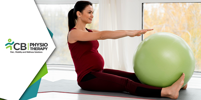
Best Exercises During Pregnancy For Each Trimester

Exercise during pregnancy is generally safe for most women and can have many benefits for both the mother and the baby. Regular physical activity can help improve mood, increase energy levels, prevent excessive weight gain, and improve sleep. Additionally, exercise during pregnancy can also improve cardiovascular fitness, muscle strength, and prepare the body for the physical demands of labor and delivery. However, it's important to consult a physiotherapist before starting or continuing any exercise regimen during pregnancy, especially if you have any medical conditions or complications. In this blog, we will discuss the exercises that can be done during pregnancy along with their benefits,
Some women may need to modify their physical activity or avoid certain exercises during pregnancy. In general, low-impact activities such as walking, swimming, and stationary cycling are recommended during pregnancy. High-impact activities, such as running, and activities that have a risk of falling or abdominal trauma, such as contact sports, should be avoided.

Guideline for Exercising During different Phases of Pregnancy:

1st Trimester (0-12 weeks):

During this phase, most women have relatively normal energy levels. Light to moderate exercises such as walking, swimming, and stationary cycling are safe and can help reduce fatigue and stress. It's important to avoid high-impact activities and activities that involve jumping, bouncing, or rapid changes of direction.
During the first trimester of pregnancy, it's important to engage in physical activities that are gentle, low-impact, and safe for both you and your baby. Stretching and strengthening exercises can help to maintain good posture, reduce aches and pains, and prepare your body for the physical demands of pregnancy and childbirth. Some stretching and strengthening exercises that can be done during the first trimester of pregnancy include:

  • Pelvic curls are a great way to begin working on spinal mobility and strengthening the abdominal muscles that support your belly as it grows.
  • Bicep curls help prepare your arms for repeatedly lifting and holding your baby.
  • Kneeling push-ups, this exercise targets the core and upper body strengthening together.
  • Cat-Cow stretching exercises help to relieve lower back pain and improve posture),
  • Hamstring stretches help to relieve tightness in the back of the legs),
  • Leg Swings help to improve circulation and relieve tension in the legs),
  • Pelvic brace or Kegels exercise helps to strengthen the pelvic floor muscles, which support the uterus and help prevent urinary incontinence),
  • Squats help to strengthen the legs and prepare them for the physical demands of childbirth),
  • Arm and Shoulder strengthening exercises help to improve posture and reduce discomfort in the upper body.

2nd Trimester (13-28 weeks):

As the baby grows and the mother's body changes, it's important to continue to modify the exercise. Avoid exercises that involve lying on your back, as this can decrease blood flow to the baby. Swimming and stationary cycling are still great options, but resistance training can also be added, focusing on the upper body, legs, and core.
During the second trimester of pregnancy, it is generally safe to perform low-impact exercises, such as:

  • Incline pushups, target the shoulder, triceps, and chest.
  • Hip and knee flexor stretching exercises are done as the center of gravity changes and the belly tends to fall forward, creating shortened hip flexor muscles. 
  • Mermaid stretching exercises can also be done as the baby grows, it can start to create pressure on the diaphragm and ribs that can be painful.
  • Side-lying leg lifts help to prepare for the changing center of gravity, it’s important to get the muscles that help with balance and assist in pelvic stabilization stronger.
  • Pelvic Tilts, This exercise helps to strengthen the lower back and abdominal muscles.
  • Squats, target the legs, glutes, and core muscles.
  • Wall push-ups help to strengthen the chest, arms, and shoulder muscles.
  • Leg Lift Exercise helps strengthen the legs, hips, and lower back muscles.
  • Bridging exercise helps strengthen the glutes, hamstrings, and lower back muscles.
  • Cat-Cow Stretch exercise stretches the spine and neck muscles.
  • Hamstring Stretching exercise stretches the hamstrings, hips, and lower back.
  • Child's Pose exercise stretches the hips, back, and legs.
  • Butterfly Stretch exercise stretches the hips, inner thighs, and lower back.
  • Seated Straddle Stretch exercise stretches the inner thighs and hips.

3rd trimester (29-40 weeks):

During this phase, the baby is growing rapidly, and the mother's body is undergoing significant changes. Exercise may become more challenging, and it's important to listen to your body and adjust the intensity and duration of exercise as needed. Gentle activities such as walking and swimming can help keep the mother active and maintain good cardiovascular health. During the third trimester of pregnancy, it is important to focus on exercises that help prepare the body for labor and delivery as it's important to stay active and maintain strength and flexibility to support your changing body.
Here are some therapeutic exercises that can be done during the third trimester:

  • Diastasis recti correction exercises are done to combat the separation of the rectus abdominal muscles during pregnancy and it shows up as a bulge that runs down the midline of the abdomen,
  • Pelvic Tilts, this exercise helps strengthen your lower back and pelvic muscles.
  • Kegels, Kegels help to strengthen your pelvic floor muscles, which support your bladder, uterus, and rectum. To do Kegels, squeeze the muscles you would use to stop urinating, hold for 5-10 seconds, then relax. Repeat 10-15 times.
  • Cat-Cow Stretch, Wall Squats, and Hamstring Stretch can be continued in this phase also.
  • Leg and foot stretch exercises help to relieve cramps and improve blood circulation.
  • Pigeon Pose: Start on all fours, then bring one knee forward to your hand and the other leg behind you. Keep your back straight and hold for 30 seconds, then switch sides.
It's important to keep in mind that every woman's pregnancy is unique and always consult a physiotherapist before starting or modifying your exercise routine. Your healthcare provider can help you determine the right level of exercise for your individual needs and make any necessary adjustments as your pregnancy progresses. However, it's important to understand that the type and intensity of exercise may need to be modified throughout pregnancy to ensure the safety of both.

Exercises to avoid during pregnancy include:

Exercises that should be avoided during pregnancy include activities that have a risk of falling, such as horseback riding, ice skating, and gymnastics, as well as activities that have a risk of an abdominal injury, such as contact sports.
  • Scuba diving can lead to changes in pressure and nitrogen absorption, which can be harmful to the developing fetus.
  • Lying on your back can cause decreased blood flow to the fetus, so exercises that involve this position should be avoided.
  • Intense cardio or High-intensity cardio workouts, such as running and jumping, can cause stress on the joints and increase the risk of injury.
  • Weightlifting during pregnancy can put extra strain on the back and lead to injury.
It's also important to listen to your body and avoid overexertion, as well as stay well-hydrated before, during, and after physical activity. If you experience any unusual symptoms such as vaginal bleeding, shortness of breath, chest pain, or contractions, stop exercising and contact your healthcare provider immediately.

Exercising during pregnancy has many benefits, such as improving mood, increasing energy levels, reducing the risk of gestational diabetes, and helping with weight management. However, it's important to understand that the type and intensity of exercise may need to be modified throughout pregnancy to ensure the safety of both the mother and the baby.

Tuesday 7 February 2023

Understanding Motor Neuron Lesions And Associated Diseases

Motor neurons are a crucial component of the nervous system, responsible for transmitting signals from the brain to the muscles. However, when these neurons suffer a lesion or injury, it can lead to debilitating conditions such as Amyotrophic Lateral Sclerosis (ALS) or Spinal Muscular Dystrophy (SMD). In this blog, we will explore motor neuron lesions and their impact on the patient's life.

Motor Neuron Lesions:

Motor neuron lesions can greatly impact an individual's mobility and daily function, but with proper physiotherapy treatment, there is hope for improvement and recovery.
Lesions in motor neurons can cause problems with the control of movement and muscle function. Motor neurons are nerve cells that control muscle contractions, and damage to these cells can result in muscle weakness, paralysis, or other movement disorders. Motor neuron lesions refer to damage or destruction of the motor neurons, which are nerve cells responsible for transmitting signals from the brain and spinal cord to the muscles. This can result in muscle weakness, difficulty in movement, and loss of muscle tone. Lesions can be caused by various factors, including degenerative diseases (such as ALS or Lou Gehrig's disease), trauma, infection, exposure to toxins, and others.
Treatment for motor neuron lesions typically involves managing symptoms and slowing the progression of the disease, and may include physiotherapy, medications, and other forms of support.

Upper Motor Neuron Lesion (UMN Lesion)
Upper motor neuron diseases refer to a group of neurological conditions that affect the upper motor neurons, which are the nerve cells in the brain that control movement and coordination. Some common examples of upper motor neuron diseases include:

  • Amyotrophic lateral sclerosis (ALS)
  • Primary lateral sclerosis (PLS)
  • Progressive bulbar palsy (PBP)
  • Progressive muscular atrophy (PMA).
An upper motor lesion is a damage to the motor pathway in the central nervous system (CNS) above the level of the spinal cord. Some signs and manual tests for UMN lesions include:
  1. Weakness,
  2. Spasticity,
  3. Muscle wasting,
  4. Increased muscle tone,
  5. Hyperreflexia,
  6. Problems with coordination movement and coordination, etc.
Manual Tests that can help diagnose an Upper Motor Lesion Include:
  • Muscle tone test: Evaluates the resistance of a limb to passive movement, looking for increased muscle tone.
  • Reflex test: Tests for hyperreflexia, which is an exaggerated knee-jerk response.
  • Clonus test: Looks for rhythmic muscular contractions caused by sudden stretching.
  • Babinski sign: Tests for the presence of an abnormal reflex that involves dorsiflexion (upward movement) of the big toe and fanning of the other toes, which is a classic sign of an upper motor lesion.
These tests can help diagnose an upper motor lesion, but further tests, such as imaging studies (e.g., MRI or CT scan) or electrophysiological tests (e.g. EMG), may be necessary to confirm the diagnosis and determine the extent of the damage. Treatment options depend on the specific condition and may include medication, physiotherapy, and other supportive measures.


Lower Motor Neuron Lesion (LMN Lesion)
Lower motor neuron diseases refer to a group of disorders that affect the lower motor neurons in the spinal cord and brainstem. Some examples of lower motor neuron diseases include:
Lower motor neuron (LMN) lesions affect the anterior horn cells of the spinal cord or the cranial nerve nuclei in the brainstem. Some signs and manual tests for LMN lesions include:
  • Muscle weakness and atrophy,
  • Hypotonia (reduced muscle tone),
  • Fasciculations (twitching of individual muscle fibres),
  • Decreased or absent deep tendon reflexes (such as knee or ankle reflexes),
  • Muscle cramps or spasms, etc.
Manual tests for LMN lesions include:
Testing for muscle weakness: The strength of specific muscle groups can be tested by asking the patient to perform certain movements (e.g., lifting a limb against resistance)
Testing for hypotonia: The muscle tone can be assessed by passively moving the limb and observing the resistance offered by the muscles
Testing for fasciculations: The presence of fasciculations can be detected by visually examining the muscles or by palpation (feeling the muscle with the hands)
Testing for reflexes: Deep tendon reflexes can be tested by tapping a tendon with a reflex hammer and observing the response of the muscle.
It is important to note that these signs and tests are not specific to LMN lesions and may also occur with other conditions. A comprehensive clinical examination, imaging studies, and other tests may be necessary to confirm a diagnosis of an LMN lesion.

Physiotherapy plays a crucial role in the rehabilitation of patients with motor neuron lesions by helping to reduce symptoms, improve their mobility, and thus increase the overall quality of life.