Tuesday 18 April 2023

Empowering The Fight Against Parkinson's Disease: Join Us In Raising Awareness And Hope On Parkinson's Day 2023


Empowering The Fight Against Parkinson's Disease: Join Us In Raising Awareness And Hope On Parkinson's Day 2023

April 11th is observed annually as Parkinson's Day to raise awareness about Parkinson's disease (PD) and its impact on individuals and communities worldwide. The theme for World Parkinson’s Day 2023 is#Take6forPD, it aims to raise awareness about the importance of exercise in PD management and encourage people with PD to engage in at least six hours of physical activity per week. Parkinson's Day is an opportunity to recognize the challenges faced by those living with PD, their families, and caregivers, and to renew our commitment to finding a cure for this debilitating disease. In this blog, we will through some light on Parkinson’s disease as well as on the theme for World Parkinson’s Day 2023, #Take6forPD.


Parkinson’s disease (PD) is a chronic and progressive movement disorder that affects millions of people worldwide. It is caused by the degeneration of dopamine-producing neurons in the brain, which leads to a variety of symptoms, including tremors, stiffness, and difficulty with balance and coordination.
While there is currently no cure for PD, there are many treatments available that can help manage symptoms and improve quality of life. One of these treatments is exercise, which has been shown to be beneficial for both physical and cognitive function in people with PD.
The campaign was launched in 2018 by the Davis Phinney Foundation, a non-profit organization dedicated to improving the lives of people with Parkinson’s. The name #Take6forPD comes from the recommendation that people with PD should aim to get at least six hours of exercise per week.

So Why is Exercise so Important for People with PD?
There are several reasons:

1. It can improve physical function: Exercise can help improve balance, coordination, flexibility, and strength, all of which can be affected by PD. By improving physical function, people with PD may be able to reduce their risk of falls and other injuries.
2. It can improve cognitive function: Exercise has been shown to improve cognitive function in people with PD, including attention, memory, and processing speed. This is important because cognitive impairment is a common non-motor symptom of PD.
3. It can improve mood: Exercise has been shown to improve mood and reduce anxiety and depression in people with PD. This is important because mood disorders are also common non-motor symptoms of PD.
4. It can improve quality of life: By improving physical and cognitive function, reducing falls and injuries, and improving mood, exercise can have a positive impact on the overall quality of life for people with PD.
 
So, What types of Exercise are recommended for People with PD?
There is no one-size-fits-all answer to this question, as the type and intensity of exercise will depend on a person’s individual needs and abilities. However, some types of exercise that may be beneficial for people with PD include:

1. Aerobic exercise: This includes activities such as walking, jogging, cycling, or swimming, and can help improve cardiovascular fitness and overall physical function.
2. Strength training: This includes weightlifting or resistance band exercises, which can help improve muscle strength and endurance.
3. Balance and coordination exercises: This includes activities such as yoga, tai chi, or dance, and can help improve balance and coordination, which are often affected by PD.
4. Flexibility exercises: This includes activities such as stretching or range of motion exercises and can help improve flexibility and range of motion.

It’s important to note that people with PD should always consult a physiotherapist before starting any new exercise program and that exercise should be tailored to their individual needs and abilities.

There are many resources available to help people with PD get started with exercise, including:
#Take6forPD is an initiative that promotes the importance of exercise for people with Parkinson's disease (PD). Exercise has been shown to have many benefits for individuals living with PD, including improving motor symptoms, increasing strength and flexibility, and enhancing the overall quality of life. However, starting an exercise program can be challenging for individuals with PD, and it is important to have access to resources and support to get started. Here are some resources that can help people with PD begin exercising:

  1. Physiotherapy: Physiotherapists specialize in designing exercise programs that meet the specific needs of individuals with PD. They can evaluate an individual's abilities and limitations and develop a tailored exercise program to address their unique needs.
  2. Parkinson's exercise classes: Many communities, such as the Rock Steady Boxing program, offer Parkinson's-specific exercise classes. These classes are led by certified instructors who are trained to work with individuals with PD and can provide guidance and support as they exercise.
  3. Online resources: There are many online resources available to help people with PD get started with exercise, including instructional videos and online classes. For example, the Parkinson's Foundation offers a free exercise program called "Parkinson's Exercise Essentials" that includes instructional videos and information on how to get started with exercise.
  4. Apps: There are many apps available that can help individuals with PD track their exercise progress and provide guidance on exercises to do. For example, the Parkinson's Exercise App provides a library of exercises that are specifically designed for individuals with PD.
  5. Support groups: Support groups can be a great resource for individuals with PD who are looking to start an exercise program. They provide an opportunity to connect with others who are facing similar challenges and can offer guidance and support as individuals navigate their exercise program.

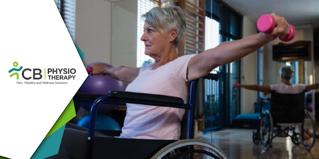
The #Take6forPD campaign is not just about raising awareness about the benefits of exercise for PD, but also about encouraging people with PD to take action and make exercise a part of their daily routine. Starting an exercise program can be challenging for individuals with PD, but with the right resources and support, it is possible to get started and experience the many benefits of exercise. By taking advantage of the resources available, individuals with PD can improve their overall health and quality of life.

Empower Your Fitness | Effective Physiotherapy Exercises For Wheelchair Users


 Empower Your Fitness | Effective Physiotherapy Exercises For Wheelchair Users

Regular exercise is important for everyone, regardless of their physical ability. However, for people who are wheelchair-bound, it can be challenging to find exercises that are safe, effective, and enjoyable. Fortunately, there are many exercises that can be done while seated in a wheelchair that can help maintain physical fitness and improve overall health. These exercises can be tailored to the specific needs and abilities of the individual and can be done in the comfort of their own home or in a physiotherapy clinic.
Before starting any Physiotherapy exercise program, it is important to consult a physiotherapist to determine what exercises are safe and appropriate for you. They can also provide guidance on how to modify exercises to accommodate any physical limitations.

Here are some exercises that are suitable for wheelchair bound individuals:
Breathing Exercises:
Breathing exercises can help improve lung capacity and reduce stress. Some examples of breathing exercises that can be done in a wheelchair include:

  • Deep breathing: The patient can take deep breaths in and out, this can help improve lung capacity and reduce stress.
  • Diaphragmatic breathing: This involves breathing from your diaphragm, which can help improve breathing efficiency and reduce shortness of breath.
  • Upper Body Strengthening Exercises:
  • These can help build and maintain strength in the arms, shoulders, and chest. Some examples of upper body strengthening exercises that can be done in a wheelchair include:
  • Bicep curls: Hold a weight in each hand and lift the weights towards your shoulders, then lower them back down.
  • Shoulder press: Hold a weight in each hand and lift the weights above your head, then lower them back down.
  • Chest press: Use resistance bands or weights to perform a chest press, pushing the weights away from your body and then bringing them back in towards your chest.
  • Wrist rotations: Rotate your wrists in a circular motion, both clockwise and counterclockwise.

Cardiovascular Exercises:
Cardiovascular exercise is important for maintaining heart health and increasing endurance. Some examples of cardiovascular exercise that can be done in a wheelchair include:
  • Wheelchair aerobics: This involves moving your arms and legs to music while sitting in a wheelchair. It can be a fun and social way to increase your heart rate.
  • Hand cycling: This involves using a handcycle machine to pedal with your arms, providing a great cardiovascular workout.
  • Wheelchair sports: Participating in sports such as wheelchair basketball, tennis, or rugby can be a fun and effective way to increase your heart rate.
Flexibility Exercises:
Flexibility exercises are important for maintaining range of motion and preventing stiffness. Some examples of flexibility exercises that can be done in a wheelchair include:
  • Seated stretches: You can perform stretches while seated in your wheelchair to improve flexibility in your upper body and legs.
  • Range of motion exercises:  ROM exercises involve moving your joints through their full range of motion to maintain flexibility.
  • Strength training:  Strength training is important for maintaining muscle mass and bone density. Some examples of strength training exercises that can be done in a wheelchair include:
  • Resistance band exercises: Resistance bands are a great way to provide resistance and build strength without the need for weights. They can be attached to the wheelchair or used with the hands and feet.
  • Weight lifting: If you have access to a gym or have weights at home, you can perform seated weightlifting exercises to build strength in your upper body. 
Balance Exercises:
Balance exercises are important for maintaining stability and preventing falls. Some examples of balance exercises that can be done in a wheelchair include:
  • Seated balance exercises: These exercises involve shifting your weight from side to side or forward and backward to improve balance.
  • Core exercises: Strengthening your core muscles can help improve balance and stability. Corestrengthening exercises can help improve posture and stability, which can reduce the risk of falls. Some examples of core strengthening exercises that can be done in a wheelchair include:
  • Seated crunches: Lean forward and bring your chest towards your knees, engaging your abdominal muscles.
  • Seated twists: Twist your torso to one side, then the other, engaging your oblique muscles.
  • Seated back extensions: Lean back in your wheelchair and engage your lower back muscles to lift your torso up.
Transfer training Exercises:
Transfer training exercises can help improve the ability to transfer in and out of a wheelchair, which can make daily activities easier and safer. Some examples of transfer training exercises that can be done in a wheelchair include:
  • Sit-to-stand transfers: Practice transferring from a seated position to a standing position, using the armrests of the wheelchair for support.
  • Bed-to-wheelchair transfers: Practice transferring from a bed to a wheelchair, using proper lifting techniques and support from a caregiver if needed.
  • Wheelchair-to-car transfers: Practice transferring from a wheelchair to a car seat, using proper techniques and support from a caregiver if needed.
These exercises can be done while seated in a wheelchair and can help maintain physical fitness and improve overall health. Consult a physiotherapist to determine what exercises are appropriate for you, and don't be afraid to try new things to keep your exercise routine interesting and engaging.

Managing Foot Drop In Spondylolisthesis | Effective Physiotherapy Techniques For Improved Mobility And Quality Of Life


Managing Foot Drop In Spondylolisthesis | Effective Physiotherapy Techniques For Improved Mobility And Quality Of Life


Foot drop and spondylolisthesis are two conditions that can have a significant impact on a person's ability to walk and carry out daily activities. While they may seem unrelated at first glance, foot drop is actually a common symptom of spondylolisthesis, a condition in which a vertebra in the spine slips out of place and onto the vertebra below it. This can cause nerve compression in the spinal canal, which can lead to a variety of symptoms, including foot drop. In this blog post, we will explore how spondylolisthesis can lead to foot drop, and how physiotherapy can help to treat these conditions.

The peroneal nerve, which controls the muscles that lift the foot, runs through the lower back and can be affected by the compression of the spinal nerves. This can cause weakness or paralysis of the muscles that control the foot, making it difficult or impossible to lift the foot upward.
In addition to nerve compression, spondylolisthesis can also cause muscle imbalances in the legs, which can contribute to foot drop. When the vertebrae in the spine shift out of place, the muscles that support the spine may become imbalanced, leading to weakness or tightness in the muscles of the legs. This can affect the way the foot is lifted and controlled, leading to foot drop.
Spondylolisthesis can also cause changes in gait and posture, which can contribute to foot drop. When a person's posture is altered due to spondylolisthesis, the muscles in the legs may be under more strain, leading to weakness or fatigue. This can affect the ability to lift the foot and control it during walking, leading to foot drop.

In some cases, foot drop may be the first sign of spondylolisthesis, alerting the patient to the presence of the condition. It is important to seek medical attention if you experience foot drop, as it may be a symptom of a more serious underlying condition There are other treatments that can help to manage the symptoms of spondylolisthesis and foot drop. These may include the use of braces or orthotics to support the affected area, medications to manage pain and inflammation, and lifestyle changes such as weight loss and exercise. In severe cases of spondylolisthesis, surgery may be necessary to relieve pressure on the nerves in the spine. This can help to alleviate symptoms such as foot drop and prevent further nerve damage. However, surgery is usually considered a last resort and is only recommended when other treatments have been ineffective.

Role of Physiotherapy
Treatment for spondylolisthesis and foot drop often involves physiotherapy to address the underlying causes of the conditions. Physiotherapy can help to improve muscle strength, flexibility, and control, as well as address any postural imbalances or gait abnormalities. This can help to alleviate the symptoms of foot drop and prevent further damage to the nerves or muscles in the affected area. Physiotherapy can be a very effective treatment for spondylolisthesis and foot drop.
In the case of spondylolisthesis, a physiotherapist can design a customized treatment plan that addresses the patient's specific needs and goals. The treatment plan may include stretching and strengthening exercises to improve the flexibility and strength of the muscles in the affected area. This can help to reduce pain and stiffness, as well as prevent further injury. For foot drop, a physiotherapist may use a variety of techniques to improve muscle function, such as electrical stimulation or gait training.

Specific Physiotherapy Techniques for Spondylolisthesis and Foot Drop
Stretching exercises can help to improve flexibility and range of motion, reducing the stiffness and pain associated with spondylolisthesis. A physiotherapist can design a program of stretching exercises that are tailored to the patient's specific needs and goals.

Strengthening exercises can help to build up the muscles around the affected area, reducing the risk of further injury and improving overall function. A physiotherapist can design a program of strengthening exercises that are appropriate for the patient's age, and fitness level.

Manual therapy techniques are also used to help alleviate the symptoms of spondylolisthesis. For example, spinal mobilization techniques can help to restore the natural movement of the vertebrae and alleviate pain and stiffness in the affected area.

Electrical stimulation can be used to activate the peroneal nerve and the muscles that control the foot. This can help to improve muscle strength and control and reduce the risk of tripping or falling. Electrical stimulation can help to activate the muscles that control the foot, thus improving the way that the patient walks, and reducing the risk of tripping or falling.

Gait training is another important aspect of physiotherapy for foot drop. This involves working with the patient to improve their gait pattern, reducing the risk of further injury, and improving overall function. A physiotherapist may use various techniques, such as mirror therapy, treadmill training, and functional electrical stimulation, to help the patient improve their gait and regain their ability to walk normally.

Education and advice are also essential components of physiotherapy for spondylolisthesis and foot drop. A physiotherapist can provide guidance on ergonomics, posture, and safe lifting techniques, helping to prevent further injury and manage symptoms. They may also provide advice on exercises that can be done at home to complement the treatment provided during physiotherapy sessions.

Spondylolisthesis and foot drop can be debilitating conditions that impact a person's ability to walk and carry out daily activities. It is important to seek treatment for spondylolisthesis and foot drop as soon as possible to prevent further damage and ensure the best possible outcome. Living with spondylolisthesis and foot drop can be challenging, but with proper treatment and management, the symptoms of both conditions can be alleviated. It is important to work closely with your physiotherapist to develop a treatment plan that addresses your specific needs and goals, helping you to regain your ability to walk normally and carry out daily activities with ease.

Thursday 6 April 2023

Get Back On Your Feet | Physiotherapist Recommended Precautions For A Successful Knee Replacement Recovery


Get Back On Your Feet | Physiotherapist Recommended Precautions For A Successful Knee Replacement Recovery


Knee replacement surgery is a major medical procedure that involves the replacement of a damaged or diseased knee joint with an artificial joint. This procedure is often recommended to patients who are suffering from severe knee pain or who have lost mobility due to arthritis or injury. After knee replacement surgery, it is crucial to follow the proper precautions and guidelines recommended by a physiotherapist to ensure a successful recovery.

The following are some essential precautions and do's and don'ts that a physiotherapist may advise after knee replacement surgery:

Follow the prescribed rehabilitation program:
After knee replacement surgery, a physiotherapist will create a customized rehabilitation program tailored to your specific needs. It is essential to follow this program diligently to ensure a smooth and successful recovery. The program may include exercises, stretches, and movements to help improve strength, flexibility, and range of motion in the knee joint.

Keep the incision site clean and dry:
It is vital to keep the incision site clean and dry to prevent infection. A physiotherapist may advise you to cover the incision site with a waterproof dressing while showering or bathing to keep it dry. Avoid submerging the incision site in water until it has completely healed.

Use assistive devices:
After knee replacement surgery, you may need to use assistive devices such as crutches, walkers, or canes to help you move around safely. A physiotherapist can help you choose the right device and provide instructions on how to use it correctly.

Manage pain and swelling:
Pain and swelling are common after knee replacement surgery. Cryotherapy (ice packs or cold compresses) can be done to reduce swelling and pain. Pain medication may also be prescribed to manage discomfort.

Avoid certain movements:
After knee replacement surgery, it is essential to avoid certain movements that can strain or damage the knee joint. A physiotherapist may advise you to avoid twisting, pivoting, or squatting on the affected leg. High-impact activities such as running or jumping should also be avoided.

Wear appropriate footwear:
Wearing appropriate footwear can help support the knee joint and prevent injuries. Shoes with good arch support and cushioning are recommended by the therapist to reduce the impact on the knee joint.

Maintain a healthy weight:
Maintaining a healthy weight can help reduce stress on the knee joint and promote a faster recovery. A physiotherapist may recommend a healthy diet and exercise program to help you maintain a healthy weight.

Gradually increase activity level:
After knee replacement surgery, it is essential to gradually increase your activity level to avoid overexerting the knee joint. Gentle exercises and a gradual increase in the intensity and duration of the activity are recommended as the knee joint heals.

Attend follow-up appointments:
It is crucial to attend all follow-up appointments with your physiotherapist and surgeon to monitor your recovery progress. These appointments may include X-rays, physical examinations, and discussions about your rehabilitation program.

Avoid driving until cleared by your surgeon:
After knee replacement surgery, it may take several weeks before you are cleared to drive. A physiotherapist may advise against driving until your surgeon has cleared you to do so.

Knee replacement surgery is a significant procedure that requires proper care and precautions to ensure a successful recovery. Following the advice of a physiotherapist is essential to reduce pain, swelling, and complications and improve mobility and function in the knee joint. By following these precautions and do's and don'ts, you can achieve a smooth and successful recovery after knee replacement surgery.

How Physiotherapy Can Relieve Lateral Knee Pain After ACL Surgery and Aid Your Recovery?


How Physiotherapy Can Relieve Lateral Knee Pain After ACL Surgery and Aid Your Recovery?


What is Lateral Knee Pain?
Lateral knee pain is a type of knee pain that is felt on the outside of the knee joint. It is often caused by an injury to the structures on the outer side of the knee, such as the iliotibial band (ITB), lateral collateral ligament (LCL), or lateral meniscus. It can also be caused by overuse or muscle imbalances in the lower extremities.

Lateral Knee Pain After ACL Reconstruction
Patients who have undergone ACL reconstruction may experience lateral knee pain as a result of the surgery. This pain can be caused by several factors, including:

  • Graft Placement: The placement of the graft during ACL reconstruction can cause lateral knee pain if it is positioned too far towards the outer side of the knee joint.
  • ITB Syndrome: The ITB can become tight and inflamed after ACL reconstruction, leading to lateral knee pain.
  • LCL Strain: The LCL can become strained during the surgery or during the rehabilitation process, leading to lateral knee pain.
  • Muscle Imbalances: Muscle imbalances in the lower extremities can cause lateral knee pain by placing excess stress on the structures on the outer side of the knee joint.
How Can Physiotherapy Help?
Physiotherapy can be an effective treatment option for patients with lateral knee pain after ACL reconstruction. The goal of physiotherapy is to reduce pain and inflammation, improve mobility, and restore strength and function to the knee joint. A qualified physiotherapist can develop a personalized treatment plan that is tailored to the patient's specific needs and goals, helping them to manage their pain and improve their quality of life.

Here are some ways that Physiotherapy can help:
  • Electrical modalities: There are several electrical modalities that can be used to relieve pain and inflammation after ACL reconstruction surgery. Here are a few examples cryotherapythermotherapyTENSUltrasound therapy, Laser therapyShockwave therapy, etc.
  • Manual Therapy: Manual therapy techniques, such as massage and joint mobilization, can help to reduce pain and inflammation in the knee joint.
  • Stretching: Stretching exercises can help to improve the flexibility of the ITB and other structures on the outer side of the knee joint, reducing stress on these structures and alleviating pain.
  • Strengthening Exercises: Strengthening exercises can help to restore strength to the muscles in the lower extremities, reducing the risk of muscle imbalances and excess stress on the knee joint.
  • Biomechanical Analysis: A physiotherapist can perform a biomechanical analysis of the patient's gait and movement patterns to identify any abnormalities that may be contributing to the lateral knee pain.
  • Bracing: A knee brace can be used to provide support to the knee joint and reduce stress on the structures on the outer side of the knee.
Lateral knee pain is a common condition that can occur after ACL reconstruction surgery. This pain can be caused by a variety of factors, including graft placement, ITB syndrome, LCL strain, and muscle imbalances. Physiotherapy can be an effective treatment option for patients with lateral knee pain, helping to reduce pain and inflammation, improve mobility, and restore strength and function to the knee joint

Genu Varum In Kids | A Physiotherapy Approach For Better Health.


Genu Varum In Kids | A Physiotherapy Approach For Better Health.


Genu varum, also known as "bow legs," is a condition in which the legs curve outward at the knees while the feet and ankles touch. While many infants are born with bow legs, the condition typically resolves on its own by age two or three. However, in some cases, the condition may persist and require medical attention, as it can cause knee pain, loss of balance, and loss of flexibility. In this blog, we will explore how parents can know if their child has genu varum and also explore how Pediatric Physiotherapy can help treat genu varum by strengthening the weak muscles and stretching the stiff muscles.

Observing the Appearance of the Legs:

One of the easiest ways to determine if your child has genu varum is to observe the appearance of their legs. Bow legs can be identified by the following physical characteristics:

1. Curved legs: If your child's legs curve outward at the knees while their feet and ankles touch, they may have bow legs.
2. Separation of the knees: When your child stands with their feet together and their knees apart, there may be a noticeable gap between the knees.
3. Tilting of the ankles: When your child stands with their feet together, their ankles may tilt inward.
4. Uneven gait: Bow legs can also affect your child's gait or the way they walk. Your child may walk with an uneven gait or have difficulty running and climbing stairs.
5. Measuring the distance between the Knees: To measure the distance between the knees, the child is asked to stand with his feet together and knees touching. A ruler or measuring tape is then placed between the knees, with the zero point at the center of the kneecaps. The distance is measured from the center of one knee to the center of the other knee. However, it's important to note that other factors, such as hip alignment and foot position, can also affect the distance between the knees.

How Physiotherapy can help treat Genu Varum in Children?

In individuals with genu varum, the tibia (shinbone) may be angled outward from the knee, while the femur (thighbone) remains straight. This can cause the muscles on the inside of the thigh (adductor muscles) to be tight, while the muscles on the outside of the thigh (abductor muscles) may be weak.
Physiotherapy can be a highly effective treatment option for individuals with genu varum. A physiotherapist will work with the patient to develop a personalized treatment plan that focuses on strengthening weak muscles, stretching stiff muscles, and improving overall alignment and stability. Here are some of the key ways that physiotherapy can help treat genu varum:

Weak muscles that need to be Strengthened:

Genu varum, the muscles that are typically weak are the abductor muscles on the outside of the thigh, and stretching alone may not be sufficient to address the muscle weakness. Stretching exercises can be beneficial for improving overall flexibility and range of motion, and may also help to reduce compensatory muscle tightness that can occur due to muscle weakness.
Muscles that are weak in children with genu varum include:
1. Gluteus medius
2. Tensor fasciae latae
These muscles are responsible for pulling the leg outward and maintaining proper alignment of the knees. Strengthening exercises, rather than stretching exercises, are typically recommended to address muscle weakness in children with genu varum.

Stiff muscles that need to be Stretched:

In addition to strengthening weak muscles, it's also important to stretch stiff muscles that may be contributing to the development of genu varum. The muscles that are tight in genu varum child and need stretching are the adductor muscles on the inside of the thigh. These include:
1. Adductor longus
2. Adductor Magnus
3. Adductor brevis
These muscles pull the leg inward and contribute to the bow-legged appearance in children with genu varum. Stretching these muscles can help to improve flexibility, reduce muscle tension, and improve the alignment of the knees.

If you suspect that your child has genu varum, it's important to consult with a physiotherapist. They can perform a physical examination and recommend an appropriate course of treatment. With the right treatment plan, most cases of genu varum can be effectively managed and corrected, allowing your child to enjoy improved stability, mobility, and overall quality of life.

Managing Genu Valgus In Children | Strengthening And Stretching Of Stiff Muscles For Improved Growth


Managing Genu Valgus In Children | Strengthening And Stretching Of Stiff Muscles For Improved Growth


Genu valgus deformity, commonly known as knock knees, is a condition where the knees come together while the feet are apart. This deformity is quite common in children and can cause various issues such as knee pain, joint pain, and difficulty in walking. Physiotherapy can be an effective treatment for genu valgus deformity in children. In this blog post, we will explore how physiotherapy can treat children with genu valgus deformity, the muscles that are weak and tight, and the exercises that can help improve the condition.

Genu valgus occurs due to a variety of reasons, including genetics, obesity, and developmental factors. During early childhood, it is normal for children to have some degree of genu valgus due to the development of the skeletal system. However, as they grow, their legs usually straighten out naturally. If genu valgus persists beyond age 7 or 8, it may indicate a problem.
Genu valgus can affect a child's growth in several ways. It can put extra stress on the knee joint and lead to pain, discomfort, and difficulty with physical activities. It can also affect their gait and balance, leading to clumsiness and an increased risk of falls. In severe cases, genu valgus can cause deformities in the legs, including bowing of the femur (thigh bone) or tibia (shin bone). This can lead to further problems with mobility and even impact the child's overall posture.
If you are concerned about your child's genu valgus, it is important to consult with a medical professional who can assess the severity of the condition and recommend appropriate treatment options. Treatment may include pediatric physiotherapy, braces, or in more severe cases, surgery.

How Does Physiotherapy Treat Genu Valgus Deformity in Children?
Physiotherapy is a non-invasive treatment option that can help improve the condition of genu valgus deformity in children. The goal of physiotherapy is to strengthen the muscles around the knee joint, improve joint stability, and correct any alignment issues. This can help improve the child's walking pattern and reduce the risk of any future knee-related problems.
The first step in physiotherapy treatment is to assess the child's condition. A physiotherapist will examine the child's gait, posture, and muscle strength. They may also take X-rays or other imaging tests to get a better understanding of the severity of the condition. Based on the assessment, the physiotherapist will develop an individualized treatment plan that may include a combination of exercises, stretches, and manual therapy techniques.

Weak Muscles in Children with Genu Valgus Deformity

Children with genu valgus deformity often have weak muscles around the hip, thigh, and knee. The following are some of the muscles that are weak in children with this condition:
  • Gluteus Medius: The gluteus medius muscle is located in the hip and is responsible for stabilizing the pelvis during walking. Weakness in this muscle can cause the pelvis to drop on the side of the affected leg, leading to an abnormal gait.
  • Vastus Medialis: The vastus medialis muscle is located on the inner side of the thigh and is responsible for extending the knee. Weakness in this muscle can cause the knee to buckle inward, exacerbating the genu valgus deformity.
  • Tibialis Anterior: The tibialis anterior muscle is located in the front of the shin and is responsible for the dorsiflexion of the foot. Weakness in this muscle can cause the foot to drop, leading to further misalignment of the knee joint.

Tight Muscles in Children with Genu Valgus Deformity

Children with genu valgus deformity often have tight muscles around the hip, thigh, and knee. The following are some of the muscles that are tight in children with this condition:
  • Adductors: The adductors are located on the inner side of the thigh and are responsible for bringing the leg toward the midline of the body. Tightness in these muscles can exacerbate the genu valgus deformity.
  • Hamstrings: The hamstrings are located on the back of the thigh and are responsible for bending the knee. Tightness in these muscles can cause the pelvis to tilt backward, leading to an abnormal gait.
  • Gastrocnemius: The gastrocnemius muscle is located in the calf and is responsible for plantarflexion of the foot. Tightness in this muscle can cause the foot to remain in a pointed position, exacerbating the genu valgus deformity.
Physiotherapy exercises can help improve the strength and flexibility of the muscles around the knee joint, leading to an improvement in the child's walking pattern.  Overall, the physiotherapist will assess the child's specific condition and prescribe a customized exercise program to address their individual needs. The goal of these exercises is to strengthen weak muscles, stretch tight muscles, and improve the overall alignment and stability of the lower extremities.

Rehabilitating Your ACL | A Physiotherapy Guide For Various Degrees Of ACL Injury

Rehabilitating Your ACL | A Physiotherapy Guide For Various Degrees Of ACL Injury

ACL (Anterior Cruciate Ligament) injury is a common type of knee injury that can range from minor sprains to complete tears. The ACL is a vital ligament that stabilizes the knee joint. When it is injured, it can cause pain, instability, and reduced function in the knee. ACL injuries are relatively common, especially among athletes, and can range from minor sprains to complete tears. Physiotherapy management for ACL injuries varies depending on the severity of the injury. In this blog, we'll look at the types of ACL injuries and how physiotherapy can help in the treatment of each type.


Grade 1 ACL Injury
A Grade 1 ACL injury is the mildest form of ACL injury, involving a minor tear or stretch in the ligament. The ligament is still intact, and the knee joint is usually stable. Physiotherapy management for a Grade 1 ACL injury focuses on reducing pain and swelling, improving the range of motion, and strengthening the surrounding muscles.

One of the primary goals of physiotherapy for a Grade 1 ACL injury is to reduce pain and inflammation. This may involve the use of ice packs, heat therapy, or electrotherapy modalities such as UltrasoundLaser therapyTENSManual therapy techniques such as soft tissue massage or joint mobilization may also be used to reduce pain and improve joint function.

The next step in physiotherapy management for a Grade 1 ACL injury is to work on restoring the normal range of motion to the knee joint. This may involve gentle stretches, active and passive range of motion exercises, and joint mobilization techniques. The physiotherapist may also use taping or bracing to help support the knee joint and prevent further injury.

Finally, strengthening exercises are an essential component of physiotherapy management for a Grade 1 ACL injury. The physiotherapist may prescribe exercises that target the muscles around the knee joint, such as the quadriceps, hamstrings, and calf muscles. These exercises may include isometric exercises, resistance training, and balance and proprioceptive exercises.

Grade 2 ACL Injury
A Grade 2 ACL injury involves a partial tear of the ligament. The knee joint may be more unstable than in a Grade 1 injury, and the patient may experience more pain and swelling. Physiotherapy management for a Grade 2 ACL injury may involve similar strategies to a Grade 1 injury, but with more emphasis on strengthening exercises to compensate for the partial tear.

In addition to the strategies used for a Grade 1 injury, the physiotherapist may prescribe more challenging strengthening exercises for a Grade 2 injury. These exercises may include eccentric strengthening exercises, which involve lengthening the muscle while it is under tension, as well as plyometric exercises, which are high-intensity exercises that involve jumping and other explosive movements.

The physiotherapist may also recommend taping or bracing to help support the knee joint and prevent further injury. In some cases, surgery may be required to repair the torn ligament, in which case physiotherapy will be an essential component of the post-operative rehabilitation process.

Grade 3 ACL injury
A Grade 3 ACL injury is the most severe form of ACL injury, involving a complete tear of the ligament. The knee joint is usually very unstable, and the patient may experience severe pain and swelling. Physiotherapy management for a Grade 3 ACL injury may involve a combination of conservative management and surgical intervention.

In the early stages of physiotherapy for a Grade 3 ACL injury, the focus is on reducing pain and swelling and regaining range of motion. The physiotherapist may use modalities such as ice packs, heat therapy, or electrotherapy to reduce pain and inflammation. Range of motion exercises and joint mobilization techniques are also essential to help maintain joint function and prevent stiffness. As the patient progresses in their rehabilitation, the physiotherapist will focus on strengthening.

After surgical intervention, physiotherapy treatment will continue to focus on restoring knee function and preventing future injuries. The rehabilitation program may include exercises to improve balance and proprioception, as well as strength and conditioning exercises to help prepare the patient for a safe return to sports or other physical activities.

Overall, the goals of physiotherapy management for ACL injuries are to reduce pain and swelling, improve range of motion and strength, and help the patient return to their pre-injury level of function. The specific strategies and exercises used will depend on the degree of ACL injury, as well as the individual needs and goals of the patient.